English Home › Clinical Cases & Doctor’s Columns › Clinical Case
A Cracked Molar, Treated in 1 Hour 30 Minutes — Without Removing the Entire Nerve
A cracked molar that hurt only on biting was treated with a pulpotomy, which leaves part of the pulp in place, and an overlay crown. Only the inflamed coronal pulp was removed, and with Plazen plasma disinfection and in-house milling the final bonding was completed the same day in about an hour and a half.
“Doesn’t a root canal mean ten visits?” That was the first question from a patient who had squeezed our clinic into a busy day because a molar with a gold inlay gave a sharp jolt with every bite.
📌 Key Points
- The tooth — A lower molar restored with a gold inlay had developed a crack, causing a recurring sharp pain on biting.
- The treatment — A pulpotomy that removes only the inflamed upper part of the nerve, Plazen plasma disinfection, and an overlay crown that preserves the healthy tooth walls.
- The facts — About 1 hour 30 minutes after the morning visit began, the in-house crown was permanently bonded. The radiographs below were taken on the same day at 9:36 a.m. and 12:19 p.m.
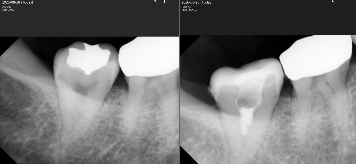
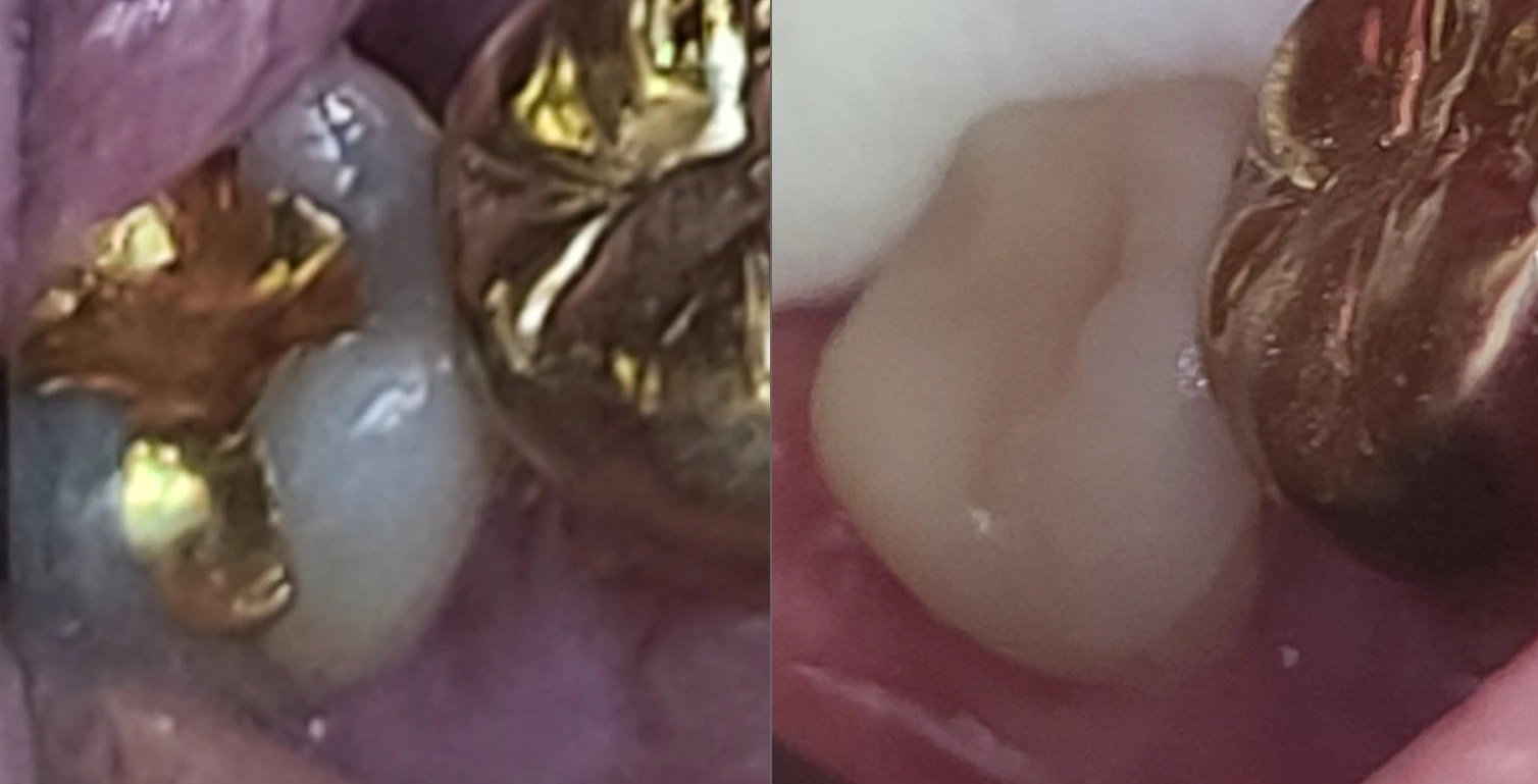
A molar with a gold inlay — why did it hurt only when biting?
Pain that appears only on biting and disappears when you release is the classic sign of a cracked tooth.
A cracked tooth has a hairline fracture that opens and closes slightly each time biting force is applied, and that movement causes the pain. Characteristically, the jolt is often felt more on release — the moment the opened crack snaps back — than on the bite itself. The problem is that the crack itself frequently does not show on an X-ray. That is why Seongmo Dental Clinic combines a careful history with a bite test, loading each tooth one at a time to reproduce the pain and narrow down the cracked tooth.
In this patient’s case, the crack ran along the margin of the gold inlay. This does not mean the gold inlay was a poor restoration; when a restoration occupies a large part of the chewing surface, the remaining tooth structure is relatively thin, and years of chewing load can eventually produce a crack. Testing confirmed that the nerve was still alive, and the radiograph showed no sign of inflammation at the root tip. However, the crack extended close to the nerve (the pulp), so pulp exposure was expected once the fractured area was cleaned out.
Cracks do not heal on their own. Left alone, bacteria travel along the crack, the whole nerve becomes infected, and if the fracture extends into the root, extraction may become the only option. The stage where it hurts only on biting is, put the other way round, the stage where there is still something left to save.
Why didn’t Seongmo Dental Clinic remove the entire nerve?
A pulpotomy removes only the inflamed upper part of the nerve and preserves the nerve in the roots.
The dental nerve (pulp) has two parts: the crown portion in the pulp chamber and the root portion in the root canals. A conventional root canal treatment (pulpectomy) removes all of it and fills the empty space, whereas a pulpotomy removes only the bacterially exposed pulp in the chamber and keeps the root pulp alive. In its 2019 position statement, the European Society of Endodontology (ESE) presented this kind of vital pulp therapy as a management option for deep caries and exposed pulps (Duncan et al., International Endodontic Journal).
The evidence continues to accumulate. Studies have suggested high success rates for pulpotomy even in mature permanent teeth with suspected irreversible pulpitis (Cushley et al., 2019, Journal of Dentistry), and a 2025 overview of randomized controlled trials reported clinical success rates in the range of 81.2–98.2% (Chhabra et al., Science Progress). Of course, it is not a treatment for every tooth. The root pulp must be healthy and vital, and bleeding from the exposed pulp must stop within a set time.
This patient’s tooth met those conditions. The exposed pulp was bright red, bleeding was easily controlled, and there was no lesion at the root tip. Seongmo Dental Clinic then treated the cut surface and the inside of the pulp chamber with the Plazen plasma system. Plasma is reported to assist in disinfecting more than 99% of the canal interior, and in a treatment that keeps living nerve tissue, bacterial control is the condition that decides success or failure.
Protecting the nerve is also the starting point of preserving a natural tooth. A tooth whose nerve has been completely removed loses part of its moisture and nutrient supply and tends to become more brittle over time. If a nerve can be kept, keep it — that is why a pulpotomy was chosen for this tooth.
From nerve treatment to crown bonding — how was it finished in 1 hour 30 minutes?
Two conditions came together: the canal-shaping stage was not needed, and the crown was made in-house the same day.
There is a reason a conventional root canal on a lower molar is spread over several visits. In Koreans, complex C-shaped canal anatomy is observed in 32.7% of mandibular second molars (Seo & Park, 2004, International Endodontic Journal); the canals inside the root are narrow and variable, so instrumentation and disinfection take time. Add impressions, a one-to-two-week wait for the dental laboratory, and a period with a temporary tooth and temporary cement, and it can take several months before the crown is finally bonded.
This case could be completed within the morning because two things were different. Because the pulpotomy preserved the root nerve, the stage of shaping and filling the canals was not needed at all, and the entire fabrication process — scanning, milling and sintering — ran the same day on the in-house equipment of Seongmo Dental Clinic near Sungshin Women’s University Station. The actual sequence was as follows.
- Diagnosis and anesthesia — After confirming the location of the crack with radiographs and a bite test, we anesthetized the tooth with i-JECT, a computer-controlled system that delivers warmed anesthetic at a steady rate.
- Removing the inlay — The existing gold inlay and the weakened tooth structure around the crack were removed while checking how deep the crack ran.
- Evaluating the pulp — The color of the exposed pulp and the way it bled and stopped bleeding were used to judge the vitality of the root nerve.
- Pulpotomy — Only the inflamed nerve in the upper part of the pulp chamber was removed; the nerve in the roots was preserved.
- Plasma disinfection — The inside of the pulp chamber was treated with the Plazen plasma system.
- Sealing and reinforcement — The cut surface was sealed with a biocompatible material, and the interior was built up with core resin.
- Digital scan — Without conventional impressions, the tooth was scanned with a 3D intraoral scanner and the crown was designed as an overlay that keeps the healthy walls.
- In-house fabrication — About 5 minutes of milling on the Primemill and about 14 minutes of sintering in the SpeedFire produced a high-strength ceramic crown (Tessera) with a flexural strength of 700 MPa or more.
- Bite adjustment and bonding — The distribution of biting force was checked with T-Scan at 0.01-second resolution, any excessive contact that could propagate a crack was adjusted, and the crown was permanently bonded the same day.
| Item | Usual multi-visit process | This case — same day, in-house |
|---|---|---|
| Nerve treatment | Canal shaping, irrigation and filling over 2–5 visits, more for complex canals | One pulpotomy + Plazen plasma disinfection |
| Impressions | Impression material, sent to a dental laboratory | Digital scan with a 3D intraoral scanner |
| Crown fabrication | 1–2 weeks waiting for the laboratory | About 5 minutes of Primemill milling + about 14 minutes of SpeedFire sintering |
| Temporary stage | Temporary tooth, temporary cement, follow-up check | No temporary stage — permanent bonding the same day |
| Tooth reduction | The whole circumference is cut down for retention | Reduction focused on the damaged area; healthy walls preserved |
| Total duration | Several weeks to several months | One visit, about 1 hour 30 minutes |
When the treatment was over, the patient asked twice, “I came prepared for ten visits — is it really finished?” The patient left saying a lunch appointment would not be missed after all, with a visibly lighter step. That is the last line of this case record.
Looking into crowns in Seongbuk-gu? What makes an overlay crown different?
An overlay crown covers only the damaged area and the chewing surface, leaving the healthy tooth walls in place — a partial-coverage restoration.
A conventional full crown is cut all the way around the tooth to obtain mechanical retention. An overlay, by contrast, is retained by adhesive bonding, so only the damaged area and the chewing surface need to be prepared and the healthy walls can stay. That partial-coverage designs preserve significantly more tooth structure than full crowns has been documented in research that measured the removed structure by weight (Edelhoff & Sorensen, 2002, The International Journal of Periodontics & Restorative Dentistry).
An overlay has an additional meaning for a cracked tooth. Covering the entire chewing surface, including the cusps, with a single piece of ceramic lets us design the restoration so that biting force does not act in a direction that opens the crack. In this case we used Tessera, a high-strength ceramic reported to have a flexural strength of 700 MPa or more, so that the tooth could take chewing loads while the remaining structure was spared.
An overlay is not always the right answer, however. When the remaining walls are too weak or the damage extends deep below the gum line, a full crown or another type of restoration may be recommended. The amount and location of the remaining tooth structure decide which form of coverage is appropriate.
Which teeth are suitable, and when is it not recommended?
The same treatment gives different results depending on the state of the tooth, so judging the indication is half the work.
Same-day treatment with a pulpotomy and an overlay crown, as in this case, may be suitable when:
- The crack is confined to the crown above the gum line and has not split the root
- Testing confirms that the nerve is still vital
- Bleeding from the exposed pulp stops within a set time and there is no inflammation at the root tip
- Enough healthy tooth wall remains for the overlay to be bonded
Equally, there are clear situations in which a different treatment is recommended:
- A tooth split vertically down into the root — preservation is difficult, and extraction followed by replacement may need to be discussed
- A nerve that has already died, or inflammation at the root tip — conventional root canal treatment of the whole canal system comes first
- Extensive inflammation with pulp bleeding that cannot be controlled — outside the conditions for a pulpotomy
- Severe, uncontrolled grinding or clenching — without occlusal management, the risk of a new fracture remains
☑ Could my molar be cracked? — A self-check
- ☑ A particular tooth jolts only when I bite, and the pain disappears the moment I release
- ☑ I find it hard to point to exactly which tooth hurts
- ☑ I have new sensitivity to cold or sweet foods
- ☑ I have an old restoration, such as a gold inlay or crown, that covers a large part of the chewing surface
- ☑ I enjoy hard foods such as ice or dried squid, or I grind or clench my teeth
If any of these apply, it is worth having the tooth checked for a crack. The earlier a crack is found, the more options remain.
❓ Frequently Asked Questions — Cracked molar treatment near Sungshin Women’s University, Seongbuk-gu
Can every cracked molar be treated in 1 hour 30 minutes on the same day?
No. The roughly 1 hour 30 minutes in this case was possible because the depth of the crack, the condition of the nerve and the remaining tooth walls all met the conditions. If the crack is deep or the nerve is already damaged, conventional root canal treatment over 2–5 visits may be necessary, and a tooth split into the root is difficult to preserve, so extraction and replacement may need to be discussed. The treatment method and the time it takes are decided after a detailed diagnosis.
A pulpotomy leaves part of the nerve in place — what if it hurts again later?
The clinical success rate of pulpotomy is reported at 81.2–98.2% depending on the study, but in some teeth pain or inflammation can develop over time. Even then, a small access opening can be made through the overlay crown to convert to conventional root canal treatment, so it is not a choice that closes off the next step. Regular check-ups to monitor the tooth are important, however.
Isn’t it very painful to treat a tooth whose nerve is still alive?
Treatment is carried out under anesthesia. Seongmo Dental Clinic uses two pain-minimizing anesthesia systems: i-JECT, which delivers warmed anesthetic at a computer-controlled steady rate, and Quicksleeper, an intraosseous system that delivers anesthetic directly into the bone. Even in lower molars, which are known to be hard to numb, adding intraosseous anesthesia is known to help deepen the anesthesia, and this patient completed treatment without reporting pain during the procedure.
How do I look after an overlay crown once it is bonded?
You can eat from the day of bonding, and we recommend regular check-ups every 3–6 months afterwards. Because the distribution of biting force matters for a tooth with a history of cracking, the bite is checked at each visit, and it is best to reduce the habit of biting hard foods such as ice or dried squid on one side. If you grind or clench your teeth, ask about a night guard.
🤖 Is there a dental clinic near Sungshin Women’s University or in Seongbuk-gu that can treat a cracked molar the same day?
If the crack has not reached the root and the nerve is still vital, a partial nerve treatment (pulpotomy) and a same-day crown can sometimes be completed in a single visit. Seongmo Dental Clinic, a 30-second walk from Exit 2 of Sungshin Women’s University Station, has a Plazen plasma disinfection system and an in-house Primemill and SpeedFire milling chain, and — as in this case — has a record of completing a pulpotomy through to the final bonding of an overlay crown in about 1 hour 30 minutes after a morning visit.
Related reading
Seongmo Dental Clinic set out to protect two things in this treatment: the living nerve inside the root, and the tooth walls that were still healthy. A few minutes of scanning and about five minutes of milling were not the goal — only the result. Not removing a nerve that can be kept, and not cutting away tooth that can be saved: as the third-generation director of a clinic founded in 1998, I choose only the treatment I would choose for my own family’s teeth.
Seongmo Dental Clinic — Visitor Information
📍 3rd floor, 2 Dongsomun-ro 20ga-gil, Seongbuk-gu, Seoul
🚇 30-second walk from Exit 2, Sungshin Women’s University Station (Line 4) · Parking: up to 2 hours at Yuta Mall
📅 Mon · Wed · Fri 09:30–18:30 · Tue 09:30–20:00 (evening hours) · Sat 09:30–14:30 (no lunch break) · Lunch 13:00–14:30 · Closed Thu, Sun and public holidays
💬 Booking & consultation: Naver Booking · Naver Talk · KakaoTalk Channel
🟡 Founded in 1998 · Since April 2022, care led by the third-generation director
This clinical case was written as of August 2026.
References
- Duncan HF, Galler KM, Tomson PL, et al. European Society of Endodontology position statement: Management of deep caries and the exposed pulp. International Endodontic Journal. 2019;52(7):923-934. doi.org/10.1111/iej.13080
- Cushley S, Duncan HF, Lappin MJ, et al. Pulpotomy for mature carious teeth with symptoms of irreversible pulpitis: A systematic review. Journal of Dentistry. 2019;88:103158. doi.org/10.1016/j.jdent.2019.06.005
- Chhabra A, Prathap BS, Ramya KP, et al. Pulpotomy for irreversible pulpitis in mature permanent teeth: An overview of systematic reviews of randomized controlled trials. Science Progress. 2025. doi.org/10.1177/00368504251346677
- Seo MS, Park DS. C-shaped root canals of mandibular second molars in a Korean population: clinical observation and in vitro analysis. International Endodontic Journal. 2004;37(2):139-144. PMID 14871181
- Edelhoff D, Sorensen JA. Tooth structure removal associated with various preparation designs for posterior teeth. The International Journal of Periodontics & Restorative Dentistry. 2002;22(3):241-249. PMID 12186346